Neurons are highly polarized cells receiving information at their dendrites while transmitting information via a single axon. Thus neurons are faced with finding the dendrites of their matching partner by axonal growth and navigating it through the nervous system and towards their target. Dendrites instead have to establish a sufficiently large surface for the many incoming and contacting axons and they do so by developing a branched tree-like network.
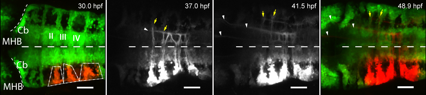
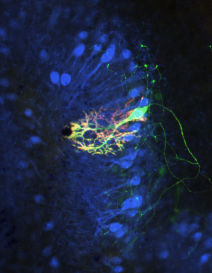
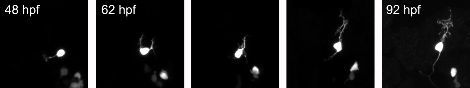
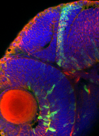
In order to better understand the function of the cerebellar system, we are trying to monitor the terminal differentiation of cerebellar neurons and in particular their developing neuronal projections. In vivo time-lapse imaging of axon and dendrite outgrowth in the transparent larval zebrafish cerebellum combined with anterograde and retrograde tracing of neuronal projections help us to elucidate the zebrafish cerebellar circuitry. Strikingly, the connections of the cerebellum in zebrafish very closely resemble the connectivity of the human cerebellum demonstrating the high conservation of this brain compartment during evolution.
Neurons pass information across their own body from dendrites to axons by differences in their electrical membrane potential, which involve local changes in ion concentrations. Fluorescent in vivo reporters of changing electrical activity allow us to watch while neurons talk within the living organism. To further unravel the function of the zebrafish cerebellum we are studying the physiology of cerebellar neuronal communication by combining genetics and in vivo imaging.
In order the understand the contribution of neuronal populations and their physiological activity to distinct behaviors one has to specifically interfere with their activity during performance task.
Novel genetic tools allow to block or trigger neuronal activity using light of specific wavelengths. We employ these optogenetic tools to interrogate cerebellar circuits to better understand how the cerebellum contributes to maintain balance of the organism and to coordinate its movements.
Selected publications:
Matsui, H., Namikawa, K., Babaryka, A., Köster R. W. (2014). Functional regionalization of the teleost cerebellum analyzed in vivo. Proceedings of the National Academy of Sciences USA (PNAS) 111: 11846-11851.
Distel, M., Wullimann, M. F., Köster, R. W. (2009). Optimized Gal4-genetics for permanent gene expression mapping in zebrafish. Proceedings of the National Academy of Sciences USA (PNAS) 106: 13365-13370.
Volkmann, K., Chen, Y.-Y., Harris, M., Wullimann, M. F., Köster, R. W. (2010). The zebrafish cerebellar upper rhombic lip generates tegmental hindbrain nuclei by long-distance migration in an evolutionary conserved manner. Journal of Comparative Neurology 518: 2794-2817.