German Research Foundation (DFG) - Project number: 404568779
The brain, as a part of the central nervous system, is probably the most complex biological system, which undergoes significant changes, especially during its growth phase. Because of these complexities—both at the macroscopic and microscopic level—and the associated difficulties in experimental sampling, there are insufficient experimental investigations. In addition, age-related mechanical investigations on brain tissue that are spatially resolved are extremely rare. However, such experiments are essential to better understand the mechanical properties and functions of the brain during growth and to develop and validate numerical models. The predictive accuracy of these models depends particularly on the quality of the identified material parameters.
The objective of this research project is the development and validation of a constitutive growth model for brain tissue that characterizes alterations of the mammalian brain in both space and time. This goal will be achieved in four steps through a close interplay of experiment, modeling, and simulation. Experiments will be performed on porcine brain tissue, which displays microstructural and mechanical properties similar to those of the human brain.
An essential prerequisite for modeling is the development of methods (I) to perform appropriate tissue-level experiments. Since grey and white matter tissue will be sampled individually, the tissue specimens are relatively small. This implies that micromechanical measurement setups under a microscope have to be developed, constructed, and verified for the axial, biaxial, and triaxial experiments planned in this project. These measurements will enable accurate characterization of the axial, biaxial, and triaxial behavior of brain tissue (II).
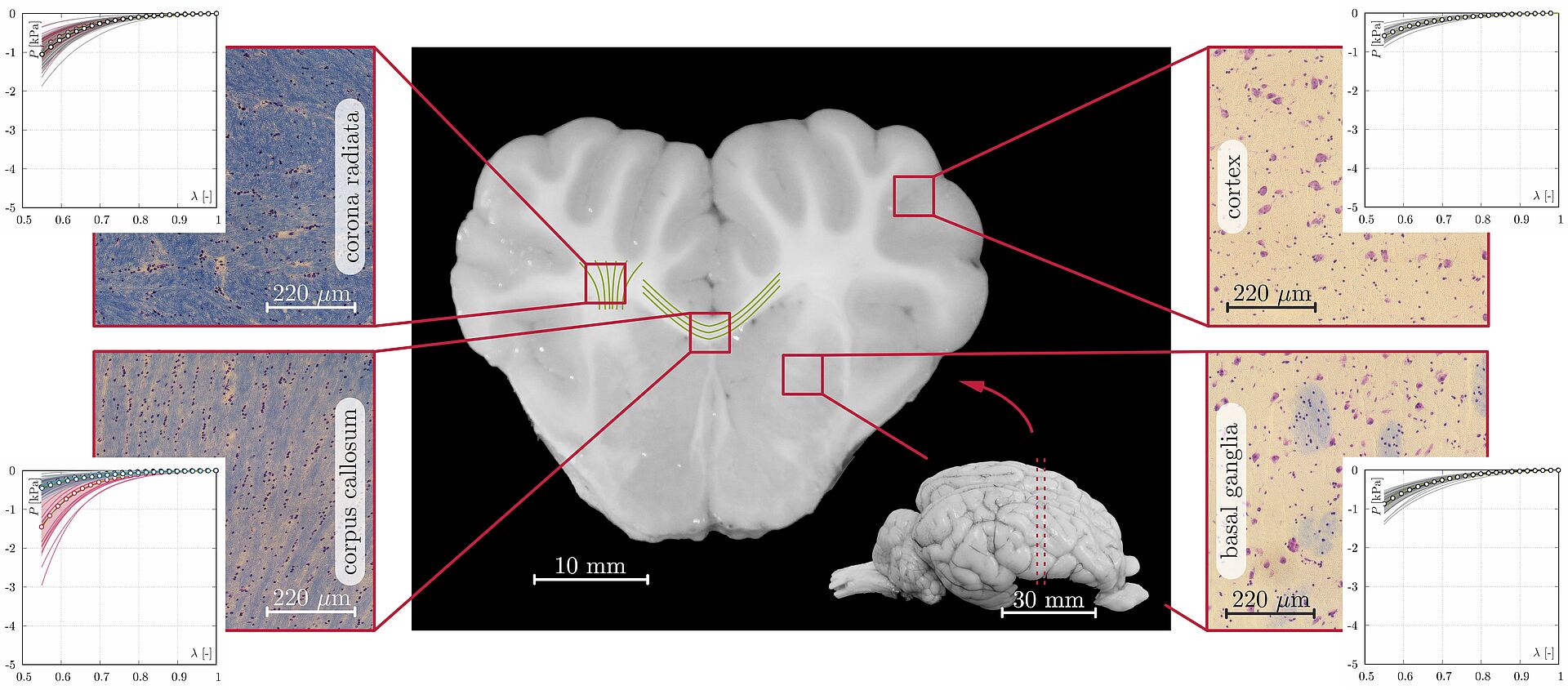
To examine the influence of neurodevelopment on mechanical properties, brain samples will be collected at different ages. In a third step, the microstructure as well as the macroscopic geometry of the brain will be determined by immunohistological investigations (III). The resulting data on cell density, myelin content, and tissue microstructure will directly inform model development and validation (IV).