
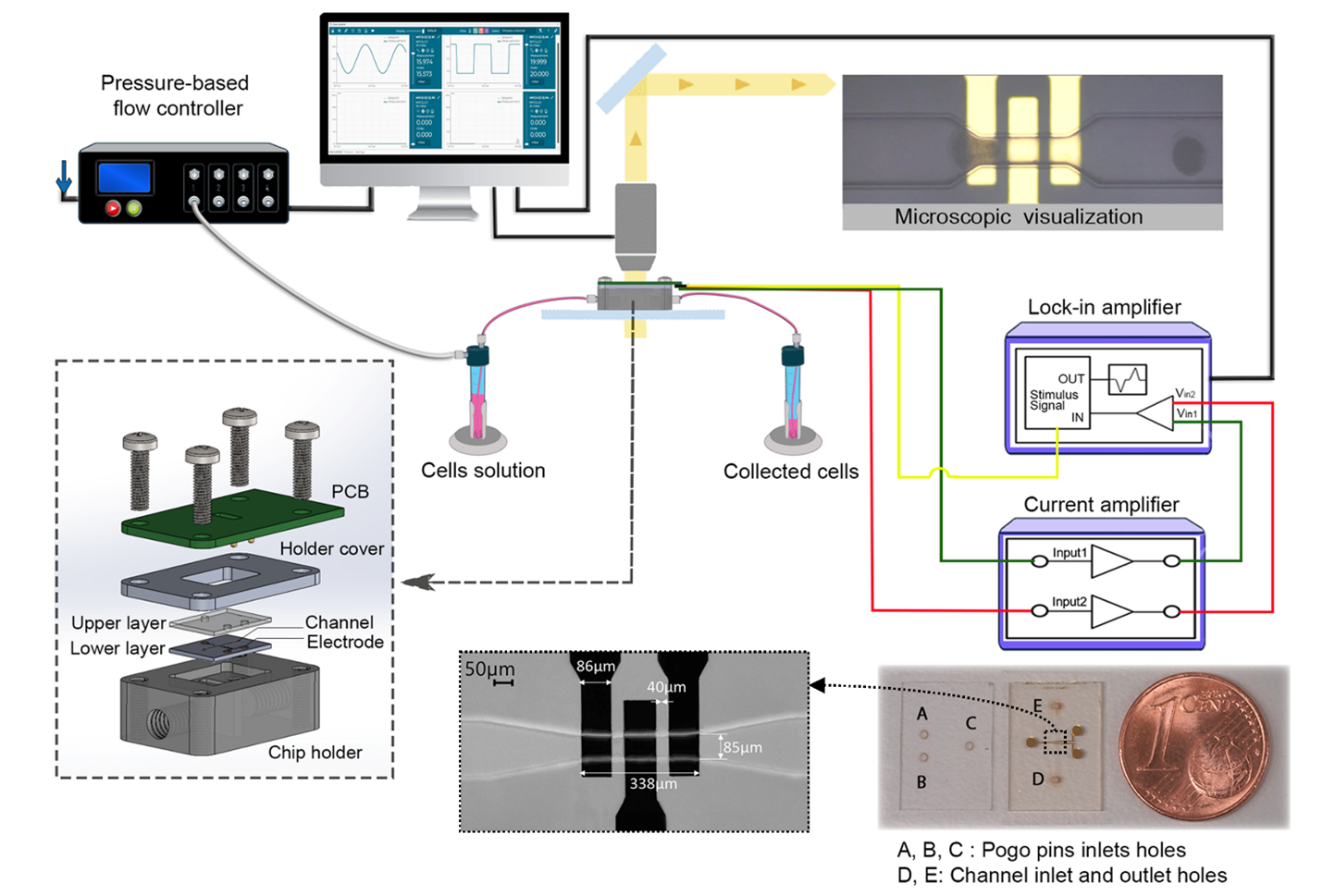
The biomechanics and dielectric properties of mammalian oocytes are key determinants of developmental competence. However, conventional approaches such as optical deformation cytometry are limited by their reliance on high-resolution imaging, requiring complex image analysis techniques and making them unsuitable for large, optically dense cells like oocytes. Here, we present a differential microfluidic impedance cytometry platform that integrates frequency-resolved dielectric profiling with constriction-based deformation analysis. The hybrid glass-SU-8 chip with integrated coplanar electrodes enables high-fidelity measurements up to 30 MHz. Using hydrogel microspheres for calibration, we confirmed that their impedance response was largely frequency-independent. In contrast, porcine oocytes displayed classical β-dispersion, reflecting membrane capacitance and cytoplasmic conductivity, as well as pressure-dependent impedance dynamics indicating viscoelastic resistance. Two-dimensional impedance mapping enabled robust discrimination between hydrogels and oocytes. In addition, fresh oocytes and oocytes recovered from a severe freeze–thaw injury model showed separable electrical and transit-dynamic signatures. Impedance-derived peak-to-valley transit (PVT) analysis notably provided a fully electrical surrogate for deformation dynamics, eliminating the need for video-based tracking. To our knowledge, this is the first application of impedance cytometry to mammalian oocytes and the first demonstration of frequency-resolved differential impedance analysis of mammalian oocytes during pressure-driven constriction transit. Compared with subjective morphological assessment and imaging-dependent deformation analysis, this approach provides objective electrical readouts of dielectric and transit-dynamic phenotypes without labelling. Uniting dielectric profiling with mechanically coupled transit metrics in a single, label-free assay enables discrimination of individual treatment-associated electromechanical phenotypes within oocyte populations. These findings establish the technical feasibility of label-free single-oocyte electromechanical phenotyping.
O. Alalul, J. Liu, M. Böl, A. Al-Halhouli, A. Dietzel
Cell-by-cell dielectric and mechanical phenotyping of oocytes
Sensing and Bio-Sensing Research, 53, 101079, (2026) [Link]